|
Article
Enamel hypoplasia on rhinocerotoid teeth: Does CT-scan imaging detect the defects better than the naked eye?
Published online: 03/01/2022
Keywords:
fossil teeth; method; micro-CT imaging; Rhinocerotoidea
https://doi.org/10.18563/pv.45.1.e2
Cited by: 5
Cite this article:
Manon Hullot and Pierre-Olivier Antoine, 2022. Enamel hypoplasia on rhinocerotoid teeth: Does CT-scan imaging detect the defects better than the naked eye?. PalaeoVertebrata 45 (1)-e2. doi: 10.18563/pv.45.1.e2
Export citation
Abstract
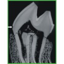
Micro-CT imaging is an increasingly popular method in paleontology giving access to internal structures with a high resolution and without destroying precious specimens. However, its potential for the study of hypoplasia defects has only recently been investigated. Here, we propose a preliminary study to test whether hypoplastic defects can be detected with micro-CT (μCT) scan and we assess the costs and benefits of using this method instead of naked eye. To do so, we studied 13 fossil rhinocerotid teeth bearing hypoplasia from Béon 1 (late early Miocene, Southwestern France) as positive control and 11 teeth of the amynodontid Cadurcotherium (Oligocene, Phosphorites du Quercy, Southwestern France), for which enamel was partly or totally obscured by cement. We showed that all macroscopically-spotted defects were retrieved on 3D reconstructions and selected virtual slices. We also detected additional defects using μCT scan compared to naked eye identification. The number of defects detected using μCT was greater in the Cadurcotherium dataset (paired-sample Wilcoxon test, p-value = 0.02724) but not for our control sample (paired-sample Wilcoxon test, p-value = 0.1171). Moreover, it allowed for measuring width and depth of the defects on virtual slices (sometimes linked to stress duration and severity, respectively), which we could not do macroscopically. As μCT imaging is both expensive and time consuming while not drastically improving the results, we recommend a moderate and thoughtful use of this method for hypoplasia investigations, restricted for instance to teeth for which enamel surface is obscured (presence of cement, uncomplete preparation, or unerupted germs).
Published in 45-1 (2022)
References
Antoine, P.-O., 2002. Phylogénie et évolution des Elasmotheriina (Mammalia, Rhinocerotidae). Mémoires du Muséum National d’Histoire Naturelle, 188:1-359.
Antoine, P.-O., Duranthon, F., 1997. Découverte de Protaceratherium minutum (Mammalia, Rhinocerotidae) dans le gisement Orléanien (MN 4) de Montréal-du-Gers (Gers). Annales de Paléontologie (Vert.-Invert.), 83:201-213.
Bacon, A.-M., Antoine, P.-O., Nguyen, T. M. H., Westaway, K., Zhao, J., Nguyen, A. T., Duringer, P., Ponche, J.-L., Sam, C. D., Truong, H. N., Tran, T. M., Nguyen, T. K. T., Pham, T. S., Demeter, S., 2020. Linear enamel hypoplasia in large-bodied mammals of Pleistocene northern Vietnam, with a special focus on Pongo. Quaternary International. https://doi.org/10.1016/j.quaint.2020.07.013
Bacon, A.-M., Antoine, P.-O., Huong, N. T. M., Westaway, K., Tuan, N. A., Duringer, P., Zhao, J., Ponche, J.-L., Dung, S. C., Nghia, T. H., Minh, T. T., Son, P. T., Boyon, M., Thuy, N. T. K., Blin, A., Demeter, F., 2018. A rhinocerotid-dominated megafauna at the MIS6-5 transition: The late Middle Pleistocene Coc Muoi assemblage, Lang Son province, Vietnam. Quaternary Science Reviews, 186:123-141. https://doi.org/10.1016/j.quascirev.2018.02.017
Barrón-Ortiz, C. I., Jass, C. N., Barrón-Corvera, R., Austen, J., Theodor, J. M., 2019. Enamel hypoplasia and dental wear of North American late Pleistocene horses and bison: an assessment of nutritionally based extinction models. Paleobiology, 45:484-515. https://doi.org/10.1017/pab.2019.17
Bratlund, B., 1999. Taubach revisited. Jahrbuch Des Römisch-Germanischen Zentralmuseums Mainz, 46:61-174.
Cabec, A. L., Tang, N., Tafforeau, P., 2015. Accessing developmental information of fossil hominin teeth using new synchrotron microtomography-based visualization techniques of dental surfaces and interfaces. PLOS ONE, 10:e0123019. https://doi.org/10.1371/journal.pone.0123019
Chollet, M. B., Teaford, M. F., 2010. Ecological stress and linear enamel hypoplasia in Cebus. American Journal of Physical Anthropology, 142:1-6. https://doi.org/10.1002/ajpa.21182
Crouzel, F., Duranthon, F., Ginsburg, F., 1988. Découverte d’un riche gisement à petits et grands mammifères d’âge Orléanien dans le département du Gers (France). Comptes Rendus de l’Académie des Sciences. Série 2, Mécanique, Physique, Chimie, Sciences de l’univers, Sciences de La Terre, 307:101-104.
Dobney, K., Ervynck, A., 1998. A protocol for recording linear enamel hypoplasia on archaeological pig teeth. International Journal of Osteoarchaeology, 8:263-273. https://doi.org/10.1002/(SICI)1099-1212(199807/08)8:4<263::AID-OA427>3.0.CO;2-P
Dobney, K., Ervynck, A., 2000. Interpreting developmental stress in archaeological pigs: the chronology of linear enamel hypoplasia. Journal of Archaeological Science, 27:597-607. https://doi.org/10.1006/jasc.1999.0477
Ensor, B. E., Irish, J. D., 1995. Hypoplastic area method for analyzing dental enamel hypoplasia. American Journal of Physical Anthropology, 98:507-517. https://doi.org/10.1002/ajpa.1330980410
Fédération Dentaire Internationale, 1982. An epidemiological index of development defects of dental enamel (DDE index). International Dental Journal, 42:411-426.
Fourvel, J.-B., Fosse, P., Fernandez, P., Antoine, P. O., 2015. Large mammals of Fouvent-Saint-Andoche (Haute-Saône, France): a glimpse into a Late Pleistocene hyena den. Geodiversitas, 37:237-266. https://doi.org/10.5252/g2015n2a5
Franco, K. M. D., Line, S. R. P., de Moura-Ribeiro, M. V. L., 2007. Prenatal and neonatal variables associated with enamel hypoplasia in deciduous teeth in low birth weight preterm infants. Journal of Applied Oral Science, 15:518-523. https://doi.org/10.1590/S1678-77572007000600012
Franz-Odendaal, T., Chinsamy, A., Lee-Thorp, J. A., 2004. High prevalence of enamel hypoplasia in an early Pliocene giraffid (Sivatherium hendeyi) from South Africa. Journal of Vertebrate Paleontology, 24:235-244. https://doi.org/10.1671/19
Franz-Odendaal, T. A., Lee-Thorp, J. A., Chinsamy, A., 2003. Insights from stable light isotopes on enamel defects and weaning in Pliocene herbivores. Journal of Biosciences, 28:765-773. https://doi.org/10.1007/BF02708437
Goodman, A. H., Rose J. C., 1990. Assessment of systemic physiological perturbations from dental enamel hypoplasias and associated histological structures. American Journal of Physical Anthropology, 33:59-110. https://doi.org/10.1002/ajpa.1330330506
Goodman, A. H., Rose J. C., 1991. Dental enamel hypoplasias as indicators of nutritional status. In: Kelley M. A., Larsen C. S. (Eds.), Advances in Dental Anthropology, Wiley-Liss, New-York, pp. 279-293.
Guatelli-Steinberg, D., 2001. What can developmental defects of enamel reveal about physiological stress in nonhuman primates? Evolutionary Anthropology: Issues, News, and Reviews, 10:138-151. https://doi.org/10.1002/evan.1027
Hassett, B. R., 2014. Missing defects? A comparison of microscopic and macroscopic approaches to identifying linear enamel hypoplasia. American Journal of Physical Anthropology, 153:463-472. https://doi.org/10.1002/ajpa.22445
Henriquez, A. C., Oxenham, M. F., 2017. An alternative objective microscopic method for the identification of linear enamel hypoplasia (LEH) in the absence of visible perikymata. Journal of Archaeological Science: Reports, 14:76-84. https://doi.org/10.1016/j.jasrep.2017.05.040
Hillman-Smith, A. K. K., Owen-Smith, N. R., Anderson, J. L., Hall-Martin, A. J., Selaladi, J. P., 1986. Age estimation of the white rhinoceros (Ceratotherium simum). Journal of Zoology, 210:355-377. https://doi.org/10.1111/j.1469-7998.1986.tb03639.x
Kierdorf, H., Witzel, C., Upex, B., Dobney, K., Kierdorf, U., 2012. Enamel hypoplasia in molars of sheep and goats, and its relationship to the pattern of tooth crown growth. Journal of Anatomy, 220:484-495. https://doi.org/10.1111/j.1469-7580.2012.01482.x
Laudet, F., Denys, C., Fernández-Jalvo, Y., 1997. Taphonomie des vertébrés oligocènes de Pech Crabit (Lot, Phosphorites du Quercy): Implications géodynamiques et paléoécologiques des remaniements post-mortem. Geobios, 30:307-313. https://doi.org/10.1016/S0016-6995(97)80036-8
Lukacs, J. R., 1999. Enamel hypoplasia in deciduous teeth of great apes: Do differences in defect prevalence imply differential levels of physiological stress? American Journal of Physical Anthropology, 110:351-363. https://doi.org/10.1002/(SICI)1096-8644(199911)110:3<351::AID-AJPA7>3.0.CO;2-2
Marchewka, J., Skrzat, J., Wrobel, A., 2014. Analysis of the enamel hypoplasia using micro-CT scanner versus classical method. Anthropologischer Anzeiger, 71:391-402. https://doi.org/10.1127/0003-5548/2014/0366
McGrath, K., El‐Zaatari, S., Guatelli-Steinberg, D., Stanton, M. A., Reid, D. J., Stoinski, T. S., Cranfield, M. R., Mudakikwa, A., McFarlin, S. C., 2018. Quantifying linear enamel hypoplasia in Virunga Mountain gorillas and other great apes. American Journal of Physical Anthropology, 166:337-352. https://doi.org/10.1002/ajpa.23436
McGrath, K., Limmer, L. S., Lockey, A.-L., Guatelli-Steinberg, D., Reid, D. J., Witzel, C., Bocaege, E., McFarlin, S. C., El Zaatari, S., 2021. 3D enamel profilometry reveals faster growth but similar stress severity in Neanderthal versus Homo sapiens teeth. Scientific Reports, 11:522. https://doi.org/10.1038/s41598-020-80148-w
Mead, A. J., 1999. Enamel hypoplasia in Miocene rhinoceroses (Teleoceras) from Nebraska: evidence of severe physiological stress. Journal of Vertebrate Paleontology, 19:391-397. https://doi.org/10.1080/02724634.1999.10011150
Ménouret, B., 2018. Le genre Cadurcotherium (Rhinocerotoidea) en Europe ; synthèse des connaissances et révision systématique. Revue de Paléobiologie, Genève, 37:495-517.
Neiburger, E. J., 1990. Enamel hypoplasias: Poor indicators of dietary stress. American Journal of Physical Anthropology, 82:231-232. https://doi.org/10.1002/ajpa.1330820211
Niven, L. B., Egeland, C. P., Todd, L. C., 2004. An inter-site comparison of enamel hypoplasia in bison: implications for paleoecology and modeling Late Plains Archaic subsistence. Journal of Archaeological Science, 31:1783-1794. https://doi.org/10.1016/j.jas.2004.06.001
Ogilvie, M. D., Curran, B. K., Trinkaus, E., 1989. Incidence and patterning of dental enamel hypoplasia among the Neandertals. American Journal of Physical Anthropology, 79:25-41. https://doi.org/10.1002/ajpa.1330790104
O’Hara, M. C., Guatelli-Steinberg, D., 2020. Differences in enamel defect expression and enamel growth variables in Macaca fascicularis and Trachypithecus cristatus from Sabah, Borneo. Journal of Archaeological Science, 114:105078. https://doi.org/10.1016/j.jas.2020.105078
Rage, J.-C., Bailón, S., 2005. Amphibians and squamate reptiles from the late early Miocene (MN 4) of Béon 1 (Montréal-du-Gers, southwestern France). Geodiversitas, 27:413-441.
Roman, F., Joleaud, L., 1909. “Cadurcotherium” de l’Isle-sur-Sorgue (Vaucluse) et révision du genre “Cadurcotherium.” Archives du Muséum d’Histoire naturelle de Lyon, 10:1-44. https://doi.org/10.3406/mhnly.1909.963
Rose, J. C., 1977. Defective enamel histology of prehistoric teeth from illinois. American Journal of Physical Anthropology, 46:439-446. https://doi.org/10.1002/ajpa.1330460309
Rothschild, B. M., Martin, L. D., Lev, G., Bercovier, H., Bar-Gal, G. K., Greenblatt, C., Donoghue, H., Spigelman, M., Brittain, D., 2001. Mycobacterium tuberculosis complex DNA from an extinct bison dated 17,000 years before the Present. Clinical Infectious Diseases, 33:305-311. https://doi.org/10.1086/321886
Sabel, N., Klingberg, G., Dietz, W., Nietzsche, S., Norén, J. G., 2010. Polarized light and scanning electron microscopic investigation of enamel hypoplasia in primary teeth. International Journal of Paediatric Dentistry, 20:31-36. https://doi.org/10.1111/j.1365-263X.2009.01006.x
Skinner, M., Goodman, A. H., 1992. Anthropological uses of developmental defects of enamel. Skeletal Biology of Past Peoples: Research Methods, 153-174.
Skinner, M. F., Hung, J. T. W., 1989. Social and biological correlates of localized enamel hypoplasia of the human deciduous canine tooth. American Journal of Physical Anthropology, 79:159-175. https://doi.org/10.1002/ajpa.1330790204
Skinner, M. F., Pruetz, J. D., 2012. Reconstruction of periodicity of repetitive linear enamel hypoplasia from perikymata counts on imbricational enamel among dry-adapted chimpanzees (Pan troglodytes verus) from Fongoli, Senegal. American Journal of Physical Anthropology, 149:468-482. https://doi.org/10.1002/ajpa.22145
Skinner, M. F., Skinner, M. M., Boesch, C., 2012. Developmental defects of the dental crown in chimpanzees from the Taï National Park, Côte d’Ivoire: coronal waisting. American Journal of Physical Anthropology, 149:272-282. https://doi.org/10.1002/ajpa.22123
Small, B. W., Murray, J. J., 1978. Enamel opacities: prevalence, classifications and aetiological considerations. Journal of Dentistry, 6:33-42. https://doi.org/10.1016/0300-5712(78)90004-0
Tafforeau, P., Bentaleb, I., Jaeger, J.-J., Martin, C., 2007. Nature of laminations and mineralization in rhinoceros enamel using histology and X-ray synchrotron microtomography: potential implications for palaeoenvironmental isotopic studies. Palaeogeography, Palaeoclimatology, Palaeoecology, 246:206-227. https://doi.org/10.1016/j.palaeo.2006.10.001
Upex, B., Dobney, K., 2012. Dental enamel hypoplasia as indicators of seasonal environmental and physiological impacts in modern sheep populations: a model for interpreting the zooarchaeological record. Journal of Zoology, 287:259-268. https://doi.org/10.1111/j.1469-7998.2012.00912.x
Wasserstein, R. L., Lazar, N. A., 2016. The ASA statement on p-Values: Context, process, and purpose. The American Statistician, 70:129-133. https://doi.org/10.1080/00031305.2016.1154108
Wasserstein, R. L., Schirm, A. L., Lazar, N. A., 2019. Moving to a World Beyond “p < 0.05.” The American Statistician, 73:1-19. https://doi.org/10.1080/00031305.2019.1583913
Windley, Z., Weller, R., Tremaine, W. H., Perkins, J. D., 2009. Two- and three-dimensional computed tomographic anatomy of the enamel, infundibulae and pulp of 126 equine cheek teeth. Part 2: Findings in teeth with macroscopic occlusal or computed tomographic lesions. Equine Veterinary Journal, 41:441-447. https://doi.org/10.2746/042516409X391033
Witzel, C., Kierdorf, U., Schultz, M., Kierdorf, H., 2008. Insights from the inside: histological analysis of abnormal enamel microstructure associated with hypoplastic enamel defects in human teeth. American Journal of Physical Anthropology, 136:400-414. https://doi.org/10.1002/ajpa.20822
Xing, S., Guatelli-Steinberg, D., O’Hara, M., Li, J., Wei, P., Liu, W., Wu, X., 2016. Micro-CT imaging and analysis of enamel defects on the Early Late Pleistocene Xujiayao juvenile. International Journal of Osteoarchaeology, 26:935-946. https://doi.org/10.1002/oa.2504
Cited by:
Davide Conedera, Yohan Pochat-Cottilloux, Nicolas Rinder, Jerôme Adrien and Jeremy E. Martin (2023).
An anatomical reappraisal of the dwarf crocodylian
Arambourgia gaudryi
from the Eocene of Quercy (France) using CT data and its implications for the phylogeny and paleoecology of basally branching alligatoroids
. Journal of Vertebrate Paleontology. https://doi.org/10.1080/02724634.2024.2313612
Manon Hullot, Céline Martin, Cécile Blondel, Damien Becker and Gertrud E. Rössner (2024). Paleobiology and paleoecology of the woolly rhinoceros (Coelodonta antiquitatis) in Northern and Central Europe: New insights from multi-proxy data. Quaternary International. https://doi.org/10.1016/j.quaint.2024.10.005
Manon Hullot, Céline Martin, Cécile Blondel, Damien Becker and Gertrud E. Rössner (2024). Evolutionary palaeoecology of European rhinocerotids across the Oligocene–Miocene transition. Royal Society Open Science. https://doi.org/10.1098/rsos.240987
Luca Pandolfi, Alberto Collareta, Dariusz Nowakowski, Giovanni Bianucci and Lorenzo Rook (2025). New early Pliocene Rhinocerotidae findings from Tuscany (Italy) and the Pliocene rhinocerotine record in Italy. Geobios. https://doi.org/10.1016/j.geobios.2023.12.012
Panagiotis Kampouridis, Luca Pandolfi, Christina Kyriakouli, Nikolai Spassov and Madelaine Böhme (2026). Deciduous dentition and ontogenetic development of the skull and teeth of Chilotherium (Mammalia, Perissodactyla, Rhinocerotidae) from the Late Miocene of Eurasia. Fossil Record. https://doi.org/10.3897/fr.29.192018
|
PDF
S.I. Data
|