Print ISSN: 0031-0247
Online ISSN: 2274-0333
Frequency: biannual
Notidanodon tooth (Neoselachii: Hexanchiformes) in the Late Jurassic of New Zealand
stratigraphy and biochronology of Oligo-Miocene of Kazakhstan
Additions to the elasmobranch fauna from the upper Cretaceous of New Jersey (middle Maastrichtian, Navesink Formation)
Fossil snakes, Palaeocene, Itaborai, Brazil, Part I
Abstract book of the 18th Conference of the EAVP
Eocene (57) , Quercy Phosphorites (38) , Systematics (32) , Rodents (29) , Mammalia (27)
Palaeovertebrata 45-1:June 2022

|
Eocene Teleostean Otoliths, Including a New Taxon, from the Clinchfield Formation (Bartonian) in Georgia, USA, with Biostratigraphic, Biogeographic,
|
|
|

|
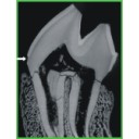
Enamel hypoplasia on rhinocerotoid teeth: Does CT-scan imaging detect the defects better than the naked eye?Manon Hullot and Pierre-Olivier AntoineKeywords: fossil teeth; method; micro-CT imaging; Rhinocerotoideadoi: 10.18563/pv.45.1.e2 Abstract Micro-CT imaging is an increasingly popular method in paleontology giving access to internal structures with a high resolution and without destroying precious specimens. However, its potential for the study of hypoplasia defects has only recently been investigated. Here, we propose a preliminary study to test whether hypoplastic defects can be detected with micro-CT (μCT) scan and we assess the costs and benefits of using this method instead of naked eye. To do so, we studied 13 fossil rhinocerotid teeth bearing hypoplasia from Béon 1 (late early Miocene, Southwestern France) as positive control and 11 teeth of the amynodontid Cadurcotherium (Oligocene, Phosphorites du Quercy, Southwestern France), for which enamel was partly or totally obscured by cement. We showed that all macroscopically-spotted defects were retrieved on 3D reconstructions and selected virtual slices. We also detected additional defects using μCT scan compared to naked eye identification. The number of defects detected using μCT was greater in the Cadurcotherium dataset (paired-sample Wilcoxon test, p-value = 0.02724) but not for our control sample (paired-sample Wilcoxon test, p-value = 0.1171). Moreover, it allowed for measuring width and depth of the defects on virtual slices (sometimes linked to stress duration and severity, respectively), which we could not do macroscopically. As μCT imaging is both expensive and time consuming while not drastically improving the results, we recommend a moderate and thoughtful use of this method for hypoplasia investigations, restricted for instance to teeth for which enamel surface is obscured (presence of cement, uncomplete preparation, or unerupted germs). Article infos |
|
S.I. Data |